
I know this is pretty heavy stuff, but its important to learn how we perceive images and color, etc. in order to fully understand anything you're creating as an art. In addition to learning a basic understanding of color and light, this in-depth information regarding the human eye and optic functions has also helped me (a beginner in photography) get a a better grasp on the concept of how the camera functions on the inside. What I have learned so far is that the human eye operates very similar to our cameras...it is an amazingly fine tuned machine.
In a basic summary from the last post, Photography and The Human Eye, the Cornea is the outermost surface of the eye, or "window" of the eye. The Iris is behind the Cornea and is like an adjustable diaphragm that controls light intensity to the Lens. The Lens is behind the Iris and adjusts to fine focus by changing shape and transmitting a focused image to the Retina in the back of the eye. The Retina the transmits the info from the Lens and through the Optic Nerve to the Brain to perceive the image.

The Retina is a highly specialized extension of the brain, which is located at the back of the eye. It contains a dense pattern of light sensitive receptor cells, called the rods and the cones. Cones can function in bright light and analyze color. While Rods provide a grey coarse vision in dim lighting.
The density of these photo receptors is also one of the things that determines the eye's ability to see in fine detail. In the central region of the retina, the rods and cones are packed even more tightly together. This part of the retina is called the Fovea and it is what gives us our sharpest vision. Away from the Fovea, the cells become more spread out, and thus making our peripheral vision the most blurred.
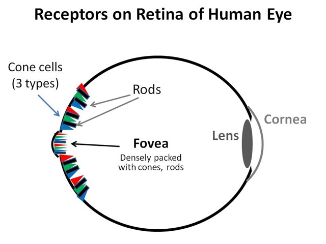
In the image below, notice the Cones (for daylight color vision) are one pattern and the Rods, which are more widely spaced, are another coarser pattern.

Image Formation
The light waves from a subject are collected and focused onto the retina by the eye's lens system. (more on light waves in the next post) Most of the refracting is done from the cornea. (more on Refraction will also be covered in the next post) The lens, which is attached at it's six edges to six muscles, focuses by changing it's shape. (See the example below)

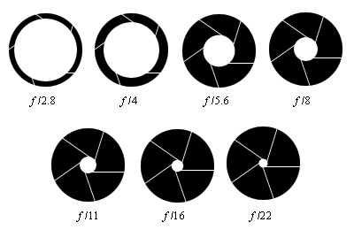
The Iris, which closely resembles an aperture ring on a camera (see aperture pic below), controls the area of the lens that is used, concentrating light waves on the more efficient central part when there is sufficient brightness. (remember, it is the Fovea in the center of the Retina that gives us the finest detail.)
The image is projected onto the retina, where photopigments from the Rods and Cones are briefly bleached to record it. This bleaching is transferred into the optic nerve and to the brain as neural activity.
(without getting too off-topic, see the neural "brain" activity pic below, which I have cropped to eliminate any confusion)

Color Sensitivity
The ability of the eye to perceive color relies on three different types of Cone receptors in the Retina, each sensitive to a different color: blue, green, and red. This trichromatic system of color receptors varies considerably in importance and frequency of use. We are least sensitive to blue wavelengths, and most sensitive to green. When all three color responses of the retina's receptors are combined, the eye's overall sensitivity is at it's peak at green-yellow. This is the reason any shade of yellow appears light (see the bananas picture below), while most blues appear dark.


Interesting bit of information: As mentioned above, humans have trichromatic vision (meaning it consists of 3 primary colors), individuals with color deficiency (color blindness) typically have the same color vision as cats and dogs, called dichromatic vision (which consists of 2 primary colors), and the most extreme color deficiency is called monochromatic vision and consists of no primary colors, only black and white. However, there are many variations of color deficiency.

Color Deficiency aka Color Blindness More In Depth
Color Deficiency is classified as a Vision Impairment, one in which impairs the ability to distinguish certain, and rarely all, colors.
Normal Cones in the Retina react to red, green, or blue when lightwaves of certain frequencies (radiating color) activate the Cones that are sensitive to the frequency. The brain interprets the varying data of intensities and blends of the Cone responses. Color deficient vision essentially means a shortage or absence of "normal" Cones to detect color the three primary colors (red, green, blue).
The most common color deficiency, which accounts for about 98% of individuals with color deficiency, is red/green deficiency, in which the person cannot distinguish red and green. A small percentage of individuals cannot distinguish blue and yellow. Rarely, a person sees only in shades of gray.
This is a pretty cool color deficiency simulator: http://www.webexhibits.org/causesofcolor/2.html
(The link would not work for some reason, so you'll have to copy and paste)
Color Deficiency Test
The most common test for color vision and color deficiency is a series of circles that contain dots of color in random patterns with a structured pattern of differing color within the field. The structured pattern may be a number (most commonly) or an object. There is no treatment to compensate for color deficiency. People who are color-deficient learn to accommodate the deficiency through mechanisms such as memorizing the locations of colored objects (such as the sequence of lights in a traffic signal) and by making adaptations in their personal environments. A person may have friends or family members sort clothing by color, for example, and label the color groups. Some people who have mild color deficiency experience benefit from devices such as colored glasses and colored contact lenses that filter the lightwaves that enter the EYE. A yellow tint may improve blue-deficient color vision, for example. This type of correction is usually only helpful for only one color.

Causes of Color Deficiency
Most color deficiency is inherited, affecting about 8 percent of men and 1⁄2 percent of women. Color deficiency may also occur following an eye injury or as a result of aging. Some other causes for example are neurological disorders such as multiple sclerosis, heavy-metal poisoning such as lead or mercury, antimalarial drugs can cause permanent changes in the retina that affect color vision, and even some erectile dysfunction medication (Viagra) can temporarily intensify the perception of blue.
No comments:
Post a Comment